# Information Form

What is Liver Cancer?
Liver cancers are malignant tumors that arise from the organ’s own tissue. See the frequency of the disease varies regionally. The prevalence of hepatitis B treatment in the past has led to the effectiveness of vaccination if significant problems arise in some countries. Hepatitis B and Hepatitis C frequent impressions in our country is a health problem. The history of the hepatitis virus is considered a risk factor that threatens the origin. Everyone who has a virus or chronic liver disease in their blood needs to be followed at regular intervals because they are at risk due to cancer.
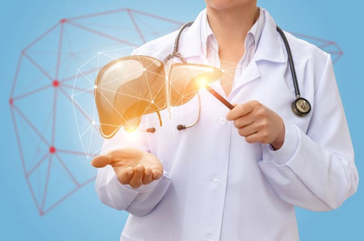
The liver is one of the largest organs in the body. It has two lobes and fills the upper right side of the abdominal cage into the rib cage. Three of the important functions of the liver are:
To filter out harmful substances from the blood can be passed through the body in the feces and urine. Safra, to help digest fat from food.
The liver is the chemical factory of medical. Storage of some nutrients. The conversion of fats into energy at the source of the body. Producing saffron, a substance that helps digestion and absorption of foods. Protein production Makes blood clotting. Production of other manufacturers that have the body’s resources. Harmful purification of alcohol, including many drugs and waste products of normal body processes. The most common type of adult primary cancer is hepatocellular carcinoma. This type of cancer is one of the third leaders in cancer-related deaths worldwide.
This summary is primary cancer. This is the summary of the treatment of cancer spreading in countries and countries in other regions of the body.
Primary cancer occurs in both adults and children. Together, treatment for children is different from treatment for adults. (For more information, see the PDQ summary on Childhood Cancer Treatment.)
The presence of hepatitis or cirrhosis may affect the risk of adult primary cancer.
Anything that increases your chances of getting sick is called a risk factor. Having a risk factor does not mean that you should get cancer; Lack of risk factors doesn’t mean you won’t get cancer. Talk to your doctor as it may be at risk.
Leading risk factor for adult primary cancer:
Have Hepatitis B or Hepatitis C. Having Hepatitis B and Hepatitis C can be even riskier.
The presence of cirrhosis can be caused by:
hepatitis (using hepatitis C); or consume alcohol for many years or become an alcoholic.
Metabolic syndrome, abdominal weight, excess fat, high blood sugar, high blood pressure, high triglyceride levels and low density in the blood, such as high lipoproteins in the form of a condition.
Liver damage lasts for a long time if it causes cirrhosis.
Having hemochromatosis is a condition in which the body receives and stores more iron from where it is heard. Extra anchored, stored in the heart and pancreas.
Eat foods stained with aflatoxin (poison from a fungus that can grow in foods such as cereals and nuts) is not stored correctly. Signs and symptoms of adult primary cancer A lump or pain on the right side.
Symptoms of liver cancer
Many patients with liver cancer do not have any symptoms in the early period. Therefore, follow-up is very important for early diagnosis, especially in high-risk patients such as cirrhosis. Liver cancers are usually abdominal distention, yellowing of the skin, itching, pain in the upper right and back of the abdomen, sudden weight loss, loss of appetite lasting for weeks, feeling of satiety and bloating after eating very little, fever, sudden worsening in general health, sweating at night darkening in the color and pale-colored stool manifests as jaundice. Although most of these symptoms are severe, they are not distinctive for liver cancer because they may all result from another condition, such as infection.
Symptoms of primary liver cancer include the following factors:
A significant amount of weight loss for an unexplained reason.
Loss of appetite for several weeks.
Don’t feel sick.
Feeling satiety and bloating after a meal, even if you eat very little.
Discomfort or pain in the abdomen.
A swollen abdomen.
Yellowish skin (jaundice), dark urine and pale stools.
Itching.
Sudden deterioration in the health of a person with chronic hepatitis or cirrhosis.
High fever and sweating. Tests examining the liver and blood are used to detect and diagnose adult primary liver cancer.
How is liver cancer diagnosed?
Blood tests: Blood tests may show abnormalities of liver function.
Imaging Tests: Your doctor may recommend imaging tests such as ultrasound, computed tomography (CT) scan, and magnetic resonance imaging (MRI).
Taking a liver tissue sample for examination. Your doctor may recommend taking a piece of liver tissue for laboratory examination to make a definitive diagnosis of liver cancer. CT scan (CAT scan): A procedure that takes detailed pictures of areas inside the body, such as the abdomen, taken from different angles. Images are made by a computer connected to an x-ray machine. A dye can be injected into a vein or swallowed to help make organs or tissues appear more clearly. This procedure is also called computed tomography, computed tomography or computed axial tomography. To obtain the best picture of abnormal areas in the liver, images can be taken at three different times after the dye is injected. This is called a three-phase CT. A spiral or helical CT scan uses an x-ray device that scans the body in a spiral path to make very detailed pictures of the areas inside the body.
MRI (magnetic resonance imaging): A procedure that uses magnets, radio waves, and computers to make detailed pictures of a number of areas within the body such as the liver. This procedure is also called nuclear magnetic resonance imaging (NMRI). To create detailed pictures of blood vessels in and near the liver, the dye is injected into a vessel. This procedure is called MRA (magnetic resonance angiography). To obtain the best picture of abnormal areas in the liver, images can be taken at three different times after the dye is injected. This is called a three-phase MRI.
Ultrasound examination: A high-energy sound waves (ultrasound) from the internal tissues or organs and echoes that form out. The echoes produced a picture of body tissues called a sonogram. The picture can be printed for later viewing.
Biopsy: The removal of cells or tissues can be seen under a microscope to check what is indicated by a pathologist on the Internet. Cell or tissue samples collection
Fine needle aspiration biopsy: Icons using a fine needle, or, or in liquid.
Nuclear Needle Biopsy: Cells or slightly larger.
Laparoscopy: A surgery to check for signs of disease to organs in the abdominal menu. Small cuts (cuts) are made on the abdominal wall and a laparoscope (thin, light tube) is placed in one. Another tool is added to remove tissue samples from the same or another incision.
Adult primary cancer does not require her time to perform a biopsy to diagnose cancer.
Some factors are prognosis (no chance of recovery) and want to treat.
Liver cancer treatment
Hepatocellular carcinoma (HCC) is the most common liver cancer and has different treatment options. Surgical treatment is the treatment method that the patients benefit most. Removal of a portion of the liver to include tumors or liver transplantation are treatment options. In the surgery, the attention is given to the fact that the remaining liver is of sufficient quality and size for the patient. Chemotherapy, radiotherapy, tumor-burning methods (ablation therapy) or microspheres and nuclear medicine treatments can be used in tumors that are not suitable for surgery or in patients who are thought to be unable to remove these large surgeries.

Karaciğer kanseri belirtileri
Karaciğer kanseri olan birçok hastada erken dönemde herhangi bir belirti olmaz.Bu nedenle özellikle siroz gibi yüksek riskli hastalarda şikayet olmasada takip erken tanı için çok önemlidir. Karaciğer kanserleri genellikle karında şişkinlik, ciltte sararma, kaşıntı, karnın sağ üst kısmından başlayıp sırta vuran ağrı, ani kilo kayıpları, haftalar süren iştahsızlık, çok az yemek yenmesine rağmen yemek sonrası tokluk ve şişkinlik hissi, ateş, geceleri terleme genel sağlıkta ani kötüleşme, idrar renginde koyulaşma ve soluk renkli dışkı gibi sarılık belirtileriyle kendini gösterir. Bu sayılan belirtilerden çoğu ağır belirtiler olsa da karaciğer kanseri demek için ayırıcı belirtiler değildir çünkü tamamı enfeksiyon gibi başka bir durumdan da kaynaklanabilir.
Primer karaciğer kanserinin belirtileri aşağıda verilen faktörleri kapsamaktadır:
Açıklanamayan nedenle önemli miktarda kilo kaybı.
Bir kaç haftalık periyodlar halinde süren iştah kaybı.
Hasta hissetme.
Çok az bir yemek olsa dahi, yemek sonrasında tokluk ve şişkinlik hissetme.
Karında (abdomen) rahatsızlık ya da ağrı.
Şişkin bir karın (abdomen).
Sarımsı cilt (sarılık), koyu renkli idrar ve soluk renkli dışkı.
Kaşıntı.
Kronik hepatit ya da siroza sahip bir kişinin sağlığındaki ani kötüleşme.
Yüksek ateş ve terleme.
Karaciğer ve kanı inceleyen testler, yetişkin birincil karaciğer kanserini tespit etmek ve teşhis etmek için kullanılır.
Karaciğer kanseri tanısı nasıl yapılır?
Kan testleri: Kan tahlilleri karaciğer fonksiyon anormalliklerini gösterebilir.
Görüntüleme Testleri: Doktorunuz, ultrason, bilgisayarlı tomografi (BT) taraması ve manyetik rezonans görüntüleme (MR) gibi görüntüleme testlerini önerebilir.
İnceleme için bir karaciğer doku örneği alınması. Doktorunuz karaciğer kanserinin kesin tanısını koyabilmek için laboratuar da incelenmek üzere bir parça karaciğer dokusunun alınmasını önerebilir. CT taraması (CAT taraması): Farklı açılardan alınan, karın gibi vücudun içindeki alanların ayrıntılı resimlerini yapan bir prosedür. Resimler bir x-ray makinesine bağlı bir bilgisayar tarafından yapılır. Bir boya, bir damar içine enjekte edilebilir veya organların veya dokuların daha net görünmesine yardımcı olmak için yutulabilir. Bu prosedüre ayrıca bilgisayarlı tomografi, bilgisayarlı tomografi veya bilgisayarlı aksiyal tomografi denir. Karaciğerdeki anormal alanların en iyi resmini elde etmek için, boya enjekte edildikten sonra görüntüler üç farklı zamanda alınabilir. Buna üç fazlı CT denir. Spiral veya sarmal CT taraması, vücudu spiral bir yolda tarayan bir röntgen cihazı kullanarak vücudun içindeki alanların çok ayrıntılı resimlerini yapar.
MRG (manyetik rezonans görüntüleme): Karaciğer gibi vücudun içindeki alanların bir dizi ayrıntılı resmini yapmak için mıknatıs, radyo dalgaları ve bilgisayar kullanan bir prosedür. Bu prosedüre ayrıca nükleer manyetik rezonans görüntüleme (NMRI) denir. Karaciğerin içinde ve yakınında kan damarlarının ayrıntılı resimlerini oluşturmak için, boya bir damar içine enjekte edilir. Bu prosedür MRA (manyetik rezonans anjiyografi) olarak adlandırılır. Karaciğerdeki anormal alanların en iyi resmini elde etmek için, boya enjekte edildikten sonra görüntüler üç farklı zamanda alınabilir. Buna üç fazlı MRI denir.
Ultrason muayenesi: Yüksek enerjili ses dalgalarının (ultrason) iç doku veya organlardan dışarı çıktığı ve ekolar oluşturduğu bir prosedür. Ekolar, sonogram adı verilen vücut dokularının bir resmini oluşturur. Resim daha sonra bakmak için basılabilir.
Biyopsi: Hücre ya da dokuların çıkarılması, böylece bir patolog tarafından kanser belirtilerini kontrol etmek için bir mikroskop altında görülebilmektedir. Hücre veya doku örneğini toplamak için kullanılan prosedürler aşağıdakileri içerir:
İnce iğne aspirasyon biyopsisi: İnce bir iğne kullanarak hücrelerin, dokunun veya sıvının alınması.
Çekirdek iğne biyopsisi: Hücrelerin veya dokunun biraz daha geniş bir iğne kullanılarak alınması.
Laparoskopi: Karın içindeki organlara hastalık belirtilerini kontrol etmek için cerrahi bir prosedür. Karın duvarında küçük kesikler (kesikler) yapılır ve insizyonlardan birine bir laparoskop (ince, ışıklı tüp) yerleştirilir. Doku numunelerini çıkarmak için aynı veya başka bir kesiden başka bir alet eklenir.
Erişkin birincil karaciğer kanserini teşhis etmek için her zaman biyopsi yapılmasına gerek yoktur.
Bazı faktörler prognozu (iyileşme şansı) ve tedavi seçeneklerini etkiler.
Karaciğer kanseri tedavisi
Hepatoselüler karsinom (HCC) en yaygın görülen karaciğer kanseridir ve farklı tedavi seçenekleri mevcuttur. Hastaların en çok yarar gördükleri tedavi yöntemi cerrahi tedavidir. Tümörleri içine alacak şekilde karaciğerin bir bölümünün çıkarılması veya karaciğer nakli tedavi seçenekleridir. Cerrahisinde dikkat edilen geriye kalacak karaciğerin hastaya yetecek nitelikte ve boyutta olmasıdır. Cerrahinin uygun olmadığı tümörlerde veya bu büyük ameliyatları kaldıramayacağı düşünülen hastalarda kemoterapi, radyoterapi, tümörün yakıldığı yöntemler (ablasyon tedavisi) veya mikroküre ile nükleer tıp tedavileri uygulanabilir.


