# Information Form

Bile duct cancer is a rare disease in which malignant cells appear in the bile ducts.
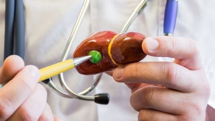
Gallbladder cancers are malignant tumors before the wall sac wall. Polyps in the gallbladder, gallbladder inflammation, obesity, nitrosamines in areas such as chemicals can cause gallbladder cancer. Those with Gall Bladder Cancer, Gall Bladder Cancer, Saffron Bladder Cancer, Developing Symptoms for Different Causes are Caught in Advanced and Advanced Stage. Small ducts come together for the right and left hepatic plants coming out of the liver. It is the region of two channels and the common hepatic channel. The common channel connects the gallbladder to the common channel. Bile hepatic ducts from the liver pass through the common hepatic duct and cystic duct and are stored in the gallbladder.
There are two types of bile duct cancer;
Intrahepatic bile duct cancer:
This type of cancers in the bile ducts in the liver are opening. Only tenacious bile duct cancer is intrahepatic. Intrahepatic bile duct cancers are also called intrahepatic cholangiocarcinomas.
Intrahepatic bile chambers are a network of small tubes carrying bile. The smallest so-called ducts come together to adjust the right hepatic bile duct and the left hepatic bile duct that drain the bile duct. It is stored in the gallbladder and the food is digested and released.
Extrahepatic bile duct cancer:
You want extrahepatic bile duct, hilum region, and a distal region. Both salvage cancer;
Perihilar bile duct cancer:
This clue is the region in the cancer hilum where the right and left bile ducts come out from the periphery and the common hepatic canal is joined. Periyar bile duct cancer is also called Klatskin tumor or perihilar cholangiocarcinoma.
Distal extrahepatic bile duct cancer:
There is such cancer called distal. The distal region from the common bile duct passing through the pancreas and ending in the small intestine. Distal extrahepatic bile duct cancer is also called extrahepatic cholangiocarcinoma.
Extrahepatic biliary chambers are small tubes that carry bile in the distant location. Common hepatic duct (hilum region) and common bile duct. Bile is put into the forest and flows into the gallbladder in the hiding place through the common hepatic and cystic channels. Bile is cut from the gallbladder by digestion.
Having colitis or clear diseases may increase the risk of bile duct cancer.
Anything that increases your risk of illness is called a risk factor. Having a risk factor does not mean that you should get cancer; Not having risk factors doesn’t mean you don’t have cancer. Those of you who think you may be at risk should discuss this with their doctor.
What are the risk factors for bile duct cancer?
– Primer sclerosing cholangitis (a progressive disease progressing with inflammation and scarring of the bile ducts)
– Chronic ulcerative colitis
– Cysts in bile ducts (cysts block bile flow and may cause swollen bile ducts, inflammation, and infection)
– Contents with Chinese liver fluke parasite.
Among the symptoms of gallbladder cancers are;
jaundice,
Abdominal pain,
Problems related to digestion
Nausea, vomiting,
Food intolerance,
Weakening,
Dark urine and white stools are observed.
Testers and dictionaries that help diagnose bile duct cancer;
Physical examination and family history:
The body’s orientation to control general symptoms. Here you will find information about the patient’s health habits, the history and the illnesses he has undergone, and the prescribed treatments.
Liver function tests:
A procedure in which the blood sample is checked to measure the amount of bilirubin and alkaline phosphatase fed by the liver to the blood. A higher amount of these substances may be a symptom of liver disease that may result from bile duct cancer.
Laboratory tests:
Medical procedures that test samples of tissue, blood, urine, or other substances in the body. These tests help diagnose, plan and control treatment or monitor the disease over time.
Carcinoembryonic antigen (CEA) and CA 19-9 tumor marker test:
A procedure in which a blood, urine or tissue sample is checked to measure the amount of certain substances made by organs, tissues, or tumor cells in the body. Some substances are found in increasing levels in the body but depend on certain types of cancer. These are called tumor markers. Higher than normal levels of carcinoembryonic antigen (CEA) and CA 19-9 may mean that they have bile duct cancer.
Ultrasound examination:
A procedure in which high-energy sound waves (ultrasound) emerge from internal tissues or organs such as the abdomen and produce echoes. Echoes form a picture of body tissues called sonograms. The picture can be printed for later viewing.
CT scan (CAT scan):
A procedure taken from different angles, detailing the areas inside the body, such as the abdomen. Images are made by a computer connected to an x-ray machine. A dye can be injected into a vein or swallowed to help make organs or tissues appear more clearly. This procedure is also called computed tomography, computed tomography or computed axial tomography.
MRI (magnetic resonance imaging):
A procedure for making detailed pictures of a number of areas inside the body using a magnet, radio wave, and a computer. This procedure is also called nuclear magnetic resonance imaging (NMRI).
MRCP (magnetic resonance cholangiopancreatography):
A procedure that uses magnets, radio waves, and computers to make detailed images of body parts such as the liver, bile ducts, gallbladder, pancreas, and pancreatic duct.
Different procedures can be used to obtain a tissue sample and diagnose bile duct cancer.
The cells and tissues are removed during a biopsy, so they can be viewed under a microscope by a pathologist to check for signs of cancer. Different procedures can be used to obtain the cell and tissue sample. The type of procedure used depends on whether the patient is sufficient for surgery.
Biopsy in Biliary Tract Cancer
Types of biopsy procedures include;
Laparoscopy:
A surgical procedure used to look at organs in the abdomen, such as the bile ducts and liver, to check for signs of cancer. Small incisions are made in the abdominal wall and a laparoscope (thin, light tube) is placed in one of the incisions. Instruments other than the same or other incisions may be added to perform procedures such as tissue samples to be checked for cancer symptoms.
Percutaneous transhepatic cholangiography (PTC):
A procedure used to x-ray the liver and bile ducts. A thin needle is inserted into the skin and liver under the ribs. The dye is injected into the liver or bile ducts and an X-ray is taken. A tissue sample is taken and checked for signs of cancer. If the bile duct becomes obstructed, a thin and flexible tube called the stent may be left in the small intestine, or the liver, to drain into the small intestine or a collection bag outside the body. This procedure can be used when a patient cannot perform surgery.
Endoscopic retrograde cholangiopancreatography (ERCP):
A procedure for x-raying ducts (tubes) carrying bile from the liver to the gallbladder and from the gallbladder to the small intestine. Sometimes bile duct cancer causes these ducts to narrow and block the flow of bile or slow down, causing jaundice. An endoscope is passed through the mouth and stomach and into the small intestine. The dye is injected into the bile ducts through an endoscope (a thin, tube-like instrument and a lens for imaging) and an x-ray is taken. A tissue sample is taken and checked for signs of cancer. If the bile duct is obstructed, a thin tube may be inserted into the duct to remove the obstruction. This tube (or stent) may be left in place to keep the duct open. This procedure can be used when a patient cannot perform surgery.
Endoscopic ultrasound (EUS):
A procedure in which an endoscope is inserted into the body, usually through the mouth or rectum. The endoscope is a thin, tube-like tool with a light and a lens to inspect. A probe at the end of the endoscope is used to remove and echo high-energy sound waves (ultrasound) from internal tissues or organs. Echoes form a picture of body tissues called sonograms. A tissue sample is taken and checked for signs of cancer. This procedure is also called endosonography.
Prognosis in Bile Cancer
Some factors affect the prognosis (chance of recovery) and treatment options.
Prognosis and treatment priority
– Tumor bile duct default
– The stage of cancer (does it only affect the bile ducts, or has it spread to the middle, lymph nodes or other places?)
– Whether cancer has spread to nearby nerves or vessels
– Can the tumor be easily removed by surgery
– If the patient’s lining is something else, such as sclerosing cholangitis
– whether the CA 19-9 level is higher than normal
– Depending on the stage of cancer or whether it has been repeated.
While treatment may be due to symptoms caused by cancer. The bile duct, the spread of cancer, subsequent examination and surgical removal can be provided by an experienced surgeon and a professional team. In this way, the adjustable lifetime and quality of life can be increased.
Diagnosis;
The methods and tests used in the diagnosis of gallbladder cancer are;
Bilirubin level and alkaline phosphatase were increased in laboratory tests. But these findings are not enough to distinguish from benign disease.
Imaging examinations such as MR, ultrasonography and/or computed tomography
A Cholangiography is used for biliary tract preparation
Angiography, ERCP (Endoscopic retrograde cholangiopancreatography) and specialized techniques such as imaging of vessels and biliary tract.
Treatment
The treatment of each case and the decision to treat is more than one, such as the location, stage, historical age and another health status of the disease. A multidisciplinary study list includes these treatments; surgery, radiotherapy, chemotherapy.
- Consultation for bile duct cancer
- The results of your imaging tests will help you to guide your process.
- Cholangiocarcinoma (bile duct cancer) treatment directs:
Whenever possible, doctors try to largely explain the description of cancer. In very small bile duct cancers, a portion of the bile duct in this section needs to be removed and the cut ends directed. In more advanced bile duct cancers, nearby forest tissue, pancreatic tissue, or lymph nodes may also be removed.
Liver transplantation. The clearance of the liver may be an option in cases where a liver donor (liver transplant) from a donor is involved in hilar cholangiocarcinoma. Although there is an improvement in hilar cholangiocarcinoma, there is no recurrence of cancer before liver transplantation.
Chemotherapy uses drugs to kill cancer cells. Before chemotherapy to a liver transplant. This can also be an option to make the patient with advanced cholangiocarcinoma ill and open beforehand and help relieve signs and symptoms.
Radiation therapy. High energy sources such as radiation chambers photon (within x) and protons are used to damage and destroy cancerous cells. A machine room (external beam radiotherapy) Or, a radioactive substance may be placed in the area close to the cancerous area within the sector (brachytherapy).
Photodynamic Therapy. To ensure the photodynamic supply, a light-sensitive chemical can be injected into a vein and collected in cancer planning to be directed. The laser light directed to cancer cells causes a chemical reaction in these cells and causes it to die. You will typically need multiple treatments. Photodynamic therapy can alleviate your signs and symptoms and at the same time slow down the growth of cancer. You should avoid direct exposure to the sun after treatments.
Bile drainage. Biliary drainage is a process in which the bile flow is restored. The cancer is able to perform a directed bypass surgery of cancer around it, or stent placement thereby keeping the bile duct collapsed due to cancer. Biliary drainage helps to alleviate the signs and symptoms of cholangiocarcinoma.